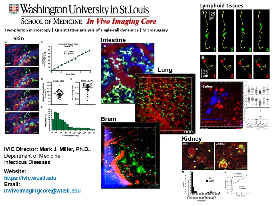
The IVIC specializes in using two-photon imaging to study disease processes at the cellular level in live mice (in vivo), human tissue specimens (ex vivo) and cell culture systems (in vitro).
Core Description
WashU School of Medicine in St. Louis offers In Vivo Imaging Core (IVIC) in Infectious Diseases to provide a cost-effective and sustainable in vivo imaging resource and multi-dimensional data analysis for WUSM researchers. The IVIC specializes in using two-photon imaging to study disease processes at the cellular level in live mice (in vivo), human tissue specimens (ex vivo) and cell culture systems (in vitro). The video below shows examples of two-photon single cell-imaging:
In Vivo Imaging Core Introductory Video
The IVIC facility is fully equipped to work with BSL2 pathogens. Users have access to microscopy rooms, wet lab and animal surgery areas, a short-term mouse holding area, a biosafety cabinet and a clean room for data analysis that has two analysis computers equipped with Imaris 9.8 software. The IVIC can assist you in designing pilot/feasibility studies, characterizing new reporter mice and analyzing your experimental data. We have expertise with small animal surgery and various imaging preparations including:
- Non-invasive in vivo imaging of skin (ear, flank, footpad) and oral mucosa.
- Intravital imaging of bowel, kidney, spleen, lymph nodes, bladder, heart, lungs, brain and spinal cord
- Imaging of ex-planted animal tissues, human biopsy specimens, cell cultures and organoids.
The IVIC provides a nexus of technical and scientific expertise for in vivo imaging methods/approaches, the development of improved methods for multi-dimensional analysis and can assists with the design and characterization of fluorescent protein reporter mouse models. A secondary goal of the IVIC is to provide hands on training opportunities for students and post-docs with interest in two-photon imaging and quantitative data analysis. The IVIC facility includes fully equipped microscope dark rooms, wet lab areas, a short-term mouse holding area and a clean room for data analysis and project consultation.
Researchers at WashU have priority for booking the equipment and services, but researchers from outside academic institutions and industry are encouraged to inquire about our services and fees for contract work. If you have any questions or would like to make a reservation or request a quote, please feel free to contact us by Email (invivoimagingcore@wustl.edu) or call (314) 362-3044.
Access
Service available to All entities, including for-profit organizations.
Priority service for Washington University only.
Services
- Two photon imaging
- Data analysis
- User Training on imaging and data analysis
Equipment
- Two-photon imaging system (1): Our two-photon microscope is a custom-made video-rate resonant scanning system built on a Olympus BX51 upright electrophysiology stand with automated x,y stage and z-focus control. The system is equipped with dual Coherent tunable femtosecond Ti-Sapphire lasers (Vision II and Chameleon XR) for efficient simultaneous excitation of both fluorescent protein reporters and traditional aromatic vital dyes, two multi-and two Bi-alkali PMTs for simultaneous 4-channel detection, Pockel’s cell optical modulators for laser attenuation and fast shuttering and high NA 20X water, 40x oil, and 20x multi-immersion media objectives.
- Acquisition computer (1) and data server (2). The two-photon imaging system has a dedicated acquisition computer which performs real-time image processing, controls the acquisition hardware and writes the multidimensional data files on a RAID 6 server/network attached storage device. Data can be acquired at 30 f/sec for a single z plane or at various time-lapse rates for a specified z-stack of images. Experimental data is stored on a 16 Tb RAID 6 NAS that is setup to tolerate two simultaneous hard drive failures for enhanced data protection. Users can transfer their data to a personal external hard drive for long-term data archiving.
- Analysis Computers and software (2). The core has two workstations running Imaris, Slidebook, Fiji, and Matlab software for 3D image rendering, cell tracking, analysis and visualization of multi-dimensional time-lapse data. Each workstation is connected to network attached storage via Gb Ethernet for data analysis and archiving. Analysis computers are also running MS Office, T-cell Analyzer and Prism for statistical analysis and generating publication ready figures, as well as Adobe Premier for editing and annotating time-lapse movies
Pricing
Pricing is subject to core verification
IVIC Access and Billing are based on a three tier system. Tier I: Contract work. All inclusive. Please contact us to discuss the scope of your project and for a cost estimate and the terms of service. Tier II: Training/fully assisted core use. This rate is $240/hr for acquisition and $160/hr for analysis and includes a staff member’s full effort for training and assisting the user during imaging preparation set up, data acquisition and cell tracking/data analysis. Tier II users must book their appointments during standard core operating hours 9 AM – 5 PM M-F. The Tier II rate does not include the cost of mice, reagents or consumable materials. You can supply your own or purchase from us. Tier III: Supervised core use. This rate is $160/hr for acquisition and $80/hr for analysis. The expectation is that Tier III users are capable of setting up their own imaging preparations, operating the equipment safely, dealing with minor technical issues and will be able to perform cell tracking and data analysis with limited trouble shooting and technical assistance by our staff. Tier III is available to users that have at least 12 hrs of fully supervised training and have passed a user proficiency test. Tier III users may book imaging and analysis time 24 hours a day 7 days a week. This rate does not include the cost of mice or consumable materials. You can supply your own or purchase from us.
AFFILIATIONS
Digestive Disease Research Core Center (DDRCC)
